

Why Full-Body Skin Exams Matter
Skin cancer is the most commonly diagnosed cancer in the United States, according to the American Academy of Dermatology (AAD). The encouraging reality is that when caught early — particularly in the case of melanoma — survival rates are significantly higher. A professional full-body skin exam is designed precisely for this purpose: systematic, trained-eye detection before a lesion becomes dangerous.
Unlike many internal cancers, skin abnormalities are visible, which makes screening both practical and highly effective. Dermatologists are trained to distinguish benign moles, seborrheic keratoses, and age spots from genuinely worrisome lesions that warrant further investigation. Understanding what happens during one of these appointments can help reduce anxiety and encourage more people to schedule them proactively.
Photograph Your Moles Between Visits
Taking dated, consistent photos of existing moles is one of the most practical tools for tracking changes between annual appointments. Store them in a dedicated album on your phone and compare them side by side at each visit. Share the photos with your dermatologist — visual documentation of change over time is clinically valuable.
What to Expect Before Your Appointment
Preparation is minimal, but a few steps make the exam more efficient and thorough.
What you will need
When you arrive, a medical assistant will typically ask about your personal and family history of skin cancer, your history of sunburns or tanning bed use, any medications that increase photosensitivity, and any spots you've noticed or that concern you. Be as specific as possible — noting when you first saw a spot and whether it has changed in size, color, or texture gives the dermatologist critical context.
How the Dermatologist Examines Your Skin
The exam itself follows a structured, head-to-toe protocol. You'll be given a gown, and the dermatologist will systematically evaluate every surface area of skin — including the scalp, behind the ears, between the toes, under the fingernails, and the soles of the feet. These less-visible areas are commonly overlooked in self-exams but are sites where skin cancers can and do develop.
Visual Inspection of the Face and Scalp
The dermatologist starts at the top, examining the face — including eyelids, lips, and ears — and then parts the hair in multiple directions to inspect the scalp. A comb or hair dryer may be used to separate hair and improve visibility. The scalp is a sun-exposed area that many patients never think to self-examine.
Examination of the Neck, Chest, and Torso
Moving downward, the clinician evaluates the neck, décolletage, chest, abdomen, and back — including the skin folds and areas that remain covered by clothing year-round. The back is one of the most common sites for melanoma in men, and it is an area almost impossible to self-examine without assistance.
Arms, Hands, and Fingernails
Each arm is examined on all sides, including the underarms (axillae) and the webbed spaces between fingers. The dermatologist will look under and around the fingernails, since a rare form of melanoma — subungual melanoma — can appear as a dark streak beneath the nail plate and is sometimes mistaken for a bruise or nail injury.
Legs, Feet, and Toenails
The dermatologist examines both legs in their entirety, including the back of the knees and inner thighs. The soles of the feet and the spaces between toes are carefully inspected. As with fingernails, toenails are checked for suspicious pigmentation. Acral lentiginous melanoma — which disproportionately affects people with darker skin tones — frequently appears on the palms and soles.
Dermoscopy for Suspicious Lesions
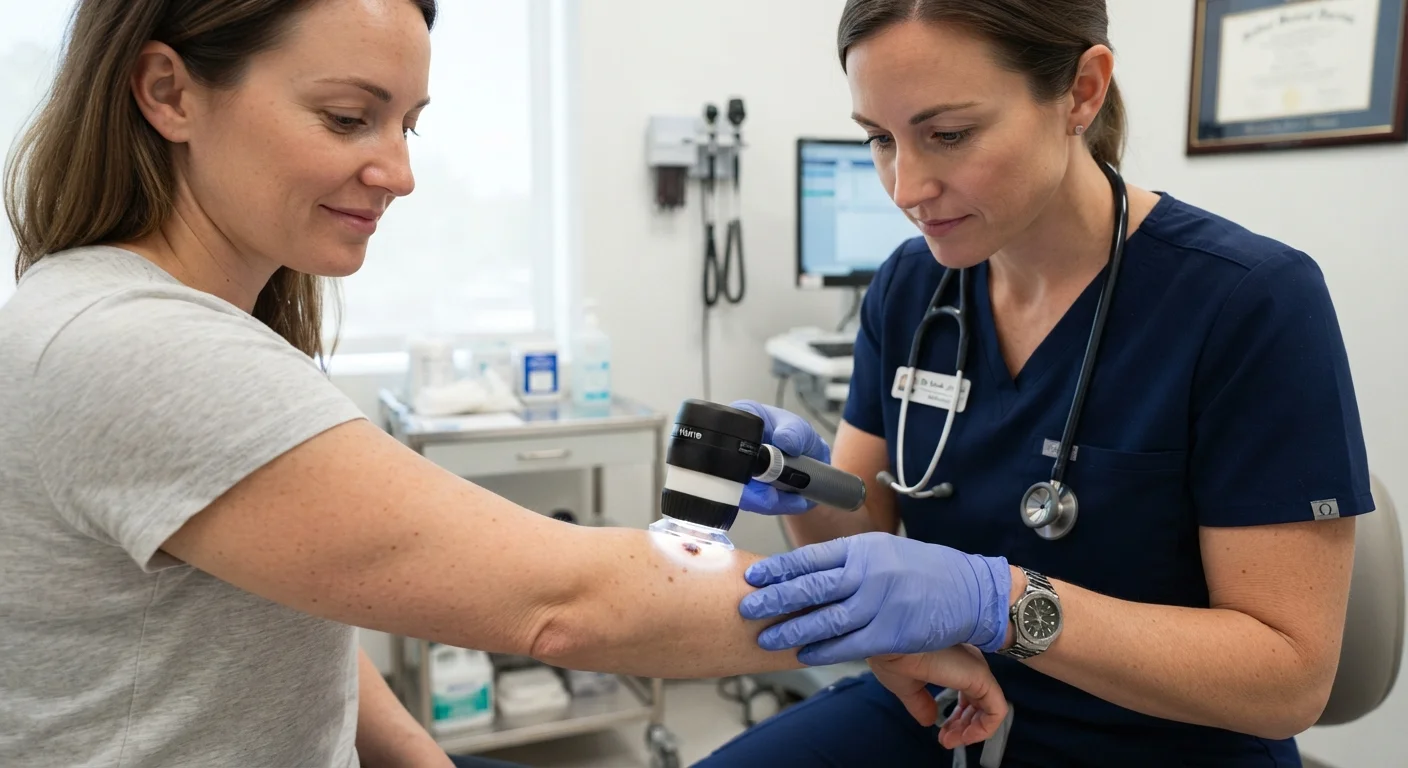
When a lesion catches the dermatologist's attention, they will examine it more closely with a dermatoscope — a handheld instrument that uses magnification and polarized light to visualize structures beneath the skin's surface invisible to the naked eye. Dermoscopy significantly improves diagnostic accuracy and reduces the number of unnecessary biopsies. The dermatologist applies the ABCDE criteria: Asymmetry, irregular Borders, multiple or unusual Colors, Diameter larger than 6 mm (about the size of a pencil eraser), and any Evolving changes in size, shape, or appearance.
Throughout the process, the dermatologist documents findings — often using a body diagram or digital photography — to track any spots over time. If a lesion looks suspicious, the next step is typically a skin biopsy, in which a small tissue sample is removed under local anesthesia and sent to a pathology lab for analysis. A biopsy is the only definitive way to diagnose skin cancer.
Who Should Get Regular Screenings and How Often
The U.S. Preventive Services Task Force (USPSTF) notes that current evidence is insufficient to assess the balance of benefits and harms of routine whole-body skin cancer screening in the general asymptomatic adult population. However, the AAD and many dermatologists recommend annual exams — or more frequent visits — for individuals with elevated risk factors.
Risk factors that may warrant more proactive screening include: a personal or family history of melanoma or other skin cancers, fair skin that burns easily, a history of significant cumulative sun exposure or indoor tanning, a large number of moles (especially atypical or dysplastic moles), and a history of organ transplantation or immunosuppression. Talk with your primary care physician or a dermatologist to determine the right screening frequency for your individual situation.
See a Dermatologist Promptly for These Signs
Do not wait for your next scheduled exam if you notice a mole or spot that is rapidly growing, bleeding without injury, itching persistently, or looks markedly different from your other moles — a concept dermatologists call the 'ugly duckling sign.' These changes can indicate a lesion that requires urgent evaluation. When in doubt, contact your dermatologist's office directly and describe what you are seeing.
Between professional exams, monthly self-examinations using the ABCDE framework are a practical complement to clinical care. Use a full-length mirror and a hand mirror for hard-to-see areas, and keep a photo log of existing moles to detect changes over time.
This article is for general informational and educational purposes only and is not a substitute for professional medical advice, diagnosis, or treatment. Always consult a qualified healthcare provider with questions about your skin health or any concerning lesions.
The content on this site is for informational purposes only and is not a substitute for professional advice. Always consult a qualified professional for guidance specific to your situation.